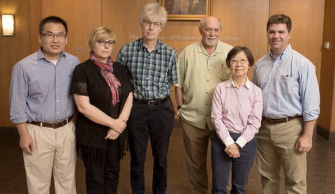
In 2013, Dmitry Vassylyev, a crystallographer at UAB, faced challenges in purifying proteins for crystallography due to the lack of a single affinity chromatography system providing ultra-high purity proteins. Existing methods suffered from flaws like sensitivity to high salt buffers, tag retention, low resin capacity, or high cost. To address this, Vassylyev devised a new approach, utilizing Colicin DNases and their inhibitors. By modifying the CE7 variant to retain binding affinity but lose toxicity, a novel CL7/Im7 system was created, where CL7 acts as a tag and Im7 as the immobilized ligand. This innovative system was detailed in a 2017 PNAS publication, offering a promising solution to the unmet need for efficient protein purification.
True Potential of Protein Purity for Structural Studies: A CLīM™ Technology Perspective
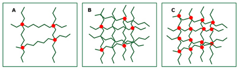
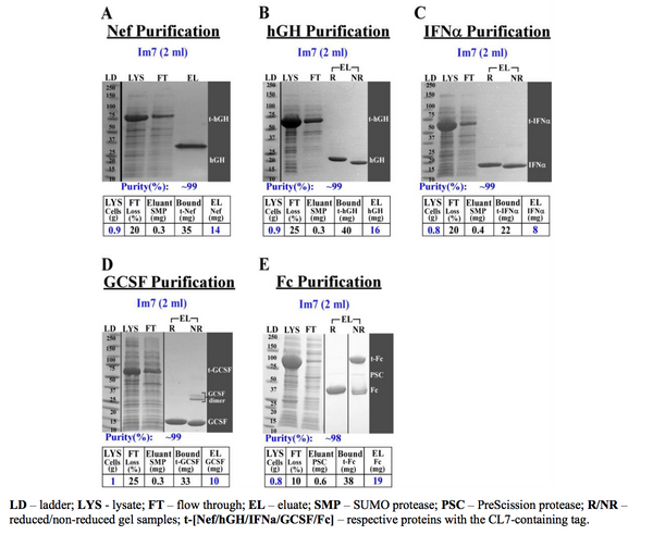
In the pursuit of protein purification, the ultimate goal is to attain a level of purity that aligns with the specific application at hand. The landscape of structural biology demands a meticulous approach, necessitating purity levels exceeding 95%. This shift is particularly pronounced in studies involving X-ray crystallography or cryo-EM, where a purity range of 95% to 99% is imperative for accurate structural insights.
Compromise on protein purity introduces inherent risks. In techniques such as X-ray crystallography, impurities hinder crystal formation, resulting in poorly diffracting crystals and low-resolution structures. Cryo-EM encounters challenges in processing images and suffers from reduced structure resolution with poorly purified proteins. Similarly, NMR spectroscopy struggles with impure proteins, making it challenging to distinguish target protein signals accurately.
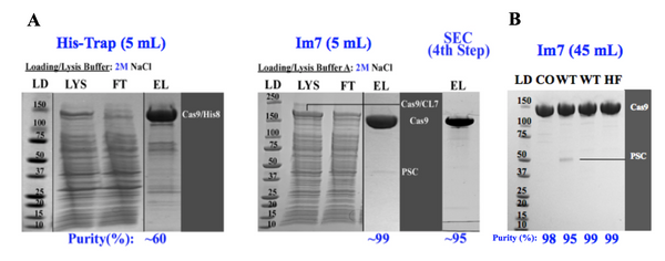
The revolutionary CLīM™ technology emerges as a streamlined solution for structural biologists. CLīM™ ensures unparalleled protein purity through a one-step chromatography process, eliminating the need for extensive multistep protocols. With its high binding capacity Im7 resin, CLīM™ technology results in efficient purification, rivaling the traditional approach of using multiple columns for different purification steps.
To achieve the pinnacle of purity for structural biology applications, TriAltus recommends the CLīM™ system over conventional chromatography methods.
TriAltus Custom Protein Purification Service for Structural Biologists:
Our custom protein purification service offers structural biologists the opportunity to delegate key projects, ensuring the highest purity and integrity of proteins for advanced structural studies. Additionally, upon request, we are fully equipped to deliver proteins in compliance with ISO 13485 standards, meeting the highest quality and regulatory requirements. Partner with TriAltus to accelerate your research while maintaining uncompromised excellence.
Contact us for a personalized consultation.
Email: info@trialtusbioscience.com
Phone: +1.205.453.8242
]]>
Here are the straightforward steps:
Both proteases are engineered for exceptional purity, speed, and efficiency. Many clients find that the minimal amount of co-eluting protease is inconsequential since the purified protein already meets their application's purity requirements.
Alternatively, a tandem affinity column can be used to capture the co-eluted protease passively as it elutes from the Im7 column.
At TriAltus, we maintain rigorous standards for assessing protein purity through two distinct methods:
These stringent assessments ensure that our protein purification processes with CLīM consistently yield high-quality and pure protein products.
TriAltus Products Needed: To implement the CLīM purification method with your CL7-tagged target protein, you'll require essential TriAltus products:
Purification protocols are also available here: (https://bit.ly/42G5d4R)
Why Choose CLīM: By opting for TriAltus' CLīM technology, you're not just purifying proteins; you're embracing a streamlined, cost-effective, high-yield method that surpasses traditional systems. Our robust one-step chromatography guarantees exceptional results.
Custom Purification Services: Unburden your team by entrusting us with your critical protein purification projects. Our custom protein purification service, powered by CLīM technology, ensures unparalleled purity and efficiency. Let us handle these tasks, allowing your team to focus on essential projects without compromising quality. We are equipped to deliver proteins in compliance with ISO 13485 standards, meeting the highest quality and regulatory requirements. Partner with TriAltus to accelerate your research while maintaining uncompromised excellence. (https://bit.ly/3HOYFrh)
Contact Us: For personalized consultations and to experience the innovation that will redefine your protein purification journey, reach out to our CLīM experts:
Email: info@trialtusbioscience.com Phone: +1.205.453.8242
]]>TriAltus Bioscience derives its name from the fusion of two Latin words: "Tri," meaning three, and "Altus," signifying high. This nomenclature is a direct representation of the company's commitment to achieving three vital benchmarks in protein purification: high purity, high yield, and high activity. These standards are not just goals but the foundation upon which TriAltus builds its innovative approaches and solutions in protein science.
The formation of TriAltus Bioscience centers around the groundbreaking CLīM (CL7/Im7) technology. Developed by researchers at the University of Alabama at Birmingham, including Professor Dmitry Vassylyev, this technology emerged from the need to overcome limitations in existing protein purification methods. The CLīM system, based on the ultra-high-affinity complex between CE7 and Im7, marked a significant advancement in the field, offering a one-step solution that achieves the coveted HHH (high-yield, high-purity, high-activity) purification.
CLīM technology is redefining the landscape of protein purification, setting a new benchmark by significantly improving the purification of complex proteins. This tag-based affinity system employs an inactive variant of CE7, named CL7, which maintains full binding affinity to Im7 without any DNA binding or DNase activity. Particularly effective for challenging biological molecules like membrane proteins, multisubunit, and DNA/RNA-binding proteins, CLīM technology excels where other methods face limitations. Unlike traditional affinity methods such as His-Trap, which can bind nonspecifically to other cellular components, the CLīM system ensures specific interaction, reducing background binding and enhancing purity. Its high capacity and stable binding in high-salt buffers, crucial for proteins with intrinsic nucleic acid-binding affinity, outperform other affinity methods in achieving the high yield, high purity, and high activity (HHH) standards. The simplicity, reusability, and broad applicability of the CLīM system make it a superior and invaluable tool in both research and industrial settings, offering a unique advantage in the purification of a diverse range of proteins.
TriAltus Bioscience stands out in the field of biotechnology for its relentless pursuit of innovation, quality, and efficiency. By embracing the CLīM technology, TriAltus has positioned itself at the forefront of protein purification science, offering products and solutions that meet the stringent requirements of today's scientific and industrial challenges. The company's dedication to maintaining the high standards encapsulated in its name is a testament to its commitment to advancing the field of protein science.
To learn more about TriAltus Bioscience and the revolutionary CLīM system, visit https://trialtusbioscience.com/. Join us as we continue to push the boundaries of protein purification technology.
]]>Welcome to the exciting world of protein purification! Today, we're going to explore the transformative power of the CLīM protein purification system. This groundbreaking technology is reshaping our approach to protein science, making purification processes more efficient, pure, and user-friendly. So, buckle up, scientists, as we embark on a journey through this revolutionary system!
At the heart of the CLīM system lies the extremely high-affinity interaction between two proteins: the CL7 tag and the small and stable immunity protein 7 (Im7) ligand. The CL7 tag is an ingeniously engineered variant of a bacterial toxin CE7 that was designed to retain ultra high affinity to the specific CE7 inhibitor, Im7, while eliminating any toxic activities. The Im7 ligand is used to make a robust specialty resin through efficient, directed coupling of the Im7 to agarose resin. Because of the extraordinary affinity, this interaction is impervious to high salt (up to 3M) and can be used at any pH between 4-10. This high-affinity pair is what makes the CLīM system an exceptional tool in our purification toolkit.
The CLīM system isn't just another method; it's a leap forward in protein purification technology. With its superior binding capacity, extraordinary stability and reusability, it stands out as a robust and economical choice for scientists. CLīM also stands out because of its high purity in a single chromatographic step, ensuring ultra high purity of the final product, and offering cost and time savings by eliminating the need for additional cleanup steps. It streamlines the purification process, delivering pure proteins efficiently and cost-effectively, making it the top value choice for scientists seeking simplicity and excellence in their research endeavors.
One of the most striking advantages of the CLīM system is its impressive binding capacity, capable of handling up to 60 mg/mL. This high capacity is not just a number; it represents a significant enhancement in purification efficiency, reducing both time and cost in your laboratory operations.
But the CL7 tag isn't all about purification. It also plays a pivotal role in boosting protein expression levels and enhancing solubility. This multifunctional aspect of the CL7 tag is particularly beneficial for complex target proteins, ensuring successful outcomes in a variety of research scenarios.
The CLīM system shines when it comes to versatility. Whether you're dealing with membrane proteins, DNA binding proteins, multisubunit complexes, or other challenging targets, CLīM adapts effortlessly, offering high-purity results even in high-salt conditions. This adaptability is a testament to the system's innovative design.
As we look to the future, the CLīM system represents a beacon of innovation in protein purification. Its potential to drive further advancements in the field is boundless, paving the way for new discoveries and developments in protein science.
In this section, we'll address some of the most common inquiries about the CL7/Im7 system, providing you with deeper insights into its capabilities and applications.
Learn more about CL7/Im7 here.
Contact us for inquiries and collaboration opportunities. info@trialtusbioscience.com
Choosing the right reagents is crucial when preparing to conduct affinity protein purification. After you’ve chosen a tag system to use, the next step is to choose an appropriate resin. This blog post will outline some of the differences between 4B and 6B agarose resin and how to choose the right resin for your needs.
Explore TriAltus’ Im7 resin products
Agarose affinity resin
Affinity chromatography resin is composed of agarose beads. Agarose beads can be used in multiple protein purification methods including size exclusion and affinity chromatography. It’s worth noting that some suppliers call their agarose “sepharose”- this is just a trademarked name for the same base material. In order to purify a protein sample using affinity chromatography, a ligand molecule is coupled to the beads. When the crude lysate is washed over the column, the ligand captures the affinity-tagged protein and remains bound while other impurities are washed away.
Differences in agarose matrices
Agarose beads come in different levels of agarose concentration which impacts several qualities of the resin. A higher concentration of agarose means that more cross-linking will occur, providing a stronger and more stable resin. This means that the resin will be more resistant to pressure buildup and can tolerate a faster flow rate. Lower agarose concentrations result in a larger pore size, while higher concentrations have a smaller pore size (Figure 1). The number of crosslinks do not affect pore size (Figure 1).
Figure 1. Agarose cross-linking and pore size at different concentrations. A- low agarose concentration, low crosslinking, B- high agarose concentration, medium crosslinking, C- high agarose concentration, high crosslinking. Source: “Introduction to Agarose Matrices,” Cube Biotech.
TriAltus Im7 affinity resins
TriAltus offers two varieties of Im7 resin: 4B and 6B. 4 and 6 refer to the % concentration of agarose in the resin sample. Both types of beads are 45-165 uM in size.
4B Resin |
6B Resin |
|
|
|
|
|
|
|
|
|
|
|
|
4B resin, because of its larger pore size, has a higher capacity of 60 mg/mL when tested with TriAltus’ 40 kDa model protein. 6B resin has about 40 mg/mL capacity. These capacity differences are preserved when a larger protein such as Cas9 (~160 kDa) is purified: 4B has 12 mg/mL capacity while 6B has 8 mg/mL. The tradeoff between the two comes down to capacity vs flow rate. Purifying large proteins with 4B resin may increase the yield due to the increased capacity and increased residence time due to slower flow rates.
In summary, 4B resin has a higher binding capacity due to larger pore size, but requires a slower flow rate. 6B resin has a slightly lower binding capacity due to its higher agarose concentration, but is less susceptible to compression and can use a faster flow rate. When used as a part of the CL7/Im7 tag system, both resin options are able to purify challenging targets.
Protein purification begins with expression in cells. Different cell expression systems offer certain benefits and drawbacks to yield and post-translational modification. TriAltus has expanded our plasmid offerings to include BacMam vectors so that the CL7/Im7 affinity purification system can be used in a wider scope of applications.
BacMam is baculovirus-mediated transduction of mammalian cells. Using a virus from insect cells (baculovirus) allows for efficient gene delivery and expression in mammalian cells, including cell types that are difficult to transfect by other means, such as primary cells or stem cells. TriAltus’ BacMam vectors don’t have selective markers to establish a stable cell line, but they can be used for transient transfection in HEK293 or CHO cells, for example.
The BacMam system works by first inserting your gene of interest into the BacMam vector and using it to generate a recombinant bacmid using the Bac-to-Bac system. This bacmid is used to transfect insect cells to generate the recombinant baculovirus that can then transduce mammalian cells.
Proteins can be expressed and purified from insect and mammalian cells separately, but BacMam combines the benefits of both options. Insect cells have high protein expression levels and allow for many of the same post-translational modifications as in mammalian cells. Insect cells also allow for the option to secrete expressed proteins into the media rather than purifying from cell lysate. Despite these benefits, some eukaryotic proteins still require expression in a mammalian cell line. BacMam bridges the gap by including mammalian transcriptional elements in a baculovirus’ DNA.
TriAltus’ BacMam vectors have both standard and unique features. Transcription is driven by a CMV promoter (human cytomegalovirus) that drives transcription in mammalian cells. In order to achieve suitable expression levels, a Kozak sequence must be included before the start codon: 5’- A/GCCATGG -3’ where ATG is the start codon. Our N-terminal tagged vector includes this sequence while the C-terminal tagged vector requires it to be added easily through your designed forward primer.
PH (polyhedron) and P10 baculovirus promoters are also included. The gene expressed by the PH promoter will be tagged, while P10 is used for co-expressing a protein if desired.
TriAltus’ BacMam vectors are unique among commercially available options in that they include three tags: CL7, His8, and eGFP. CL7 gives you the ability to purify your protein of interest using Im7 resin. His8 is a common tag for many vectors and is available for those who require double-tagging. eGFP is included for visualization purposes. All three tags are cleavable from the protein of interest using PSC protease.
BacMam NT H8-CL7-eGFP vector map
BacMam CT eGFP-CL7-H8 vector map
Download Genbank files of the BacMam vectors from our Plasmid Guide hereBacMam is a useful tool for scientists who want to combine the benefits of insect and mammalian expression systems to produce well-expressed, post-translationally modified proteins.
Endotoxin testing is an important standard of many proteins’ quality control reporting. Verifying the amount of endotoxin in a sample of protein is critical to ensure that it is safe for research and therapeutic applications.
Endotoxins are a type of pyrogen, meaning that they cause a fever response with enough volume in an organism. These lipopolysaccharides are found in the cell walls of gram-negative bacteria. It is crucial to know if the level of endotoxin present in a sample is below a safe threshold, especially for injectable pharmaceuticals and medical implants. Even for research purposes, it is important to avoid toxic materials that could confound results, especially in cell culture.
Endotoxin activity levels are reported in EU/mL (endotoxin units). One EU is roughly equivalent to 0.1-0.2 ng endotoxin/mL of solution. The FDA sets restrictions on acceptable levels in devices and non-oral drugs, but there is not a set standard for research purposes. Recombinant proteins for research tend to have <1.0 EU/ug. TriAltus’ human growth hormone has <0.1 EU/ug and Cas9 has <0.3 EU/ug as measured by the rFC method.
Endotoxin testing began with the rabbit pyrogen method in which a rabbit would be injected with the sample in question. If the rabbit showed a fever response, the sample was assumed to contain an unacceptable level of endotoxin. However, this method was too non-specific, and the LAL test quickly took hold as the standard procedure.
LAL (Limulus amoebocyte lysate) testing originates from the blood of horseshoe crabs. The presence of endotoxin in the lysate triggers proteins such as recombinant factors C, G, and B in a signaling cascade that activates a clotting enzyme. It is the cascade that allows for the extreme sensitivity of the test. To test for endotoxin, samples are mixed with LAL, and coagulation is measured either through a chromogenic or a turbidimetric assay. These tests can register up to 0.01 EU/mL.
Despite being the most widely used means of endotoxin testing, LAL has several downsides. Horseshoe crabs and their blood are highly limited resources bordering on endangered. LAL has natural variations between samples due to differences among the population of horseshoe crabs. Additionally, the clotting cascade can be initiated by off-target effects and decrease the accuracy of the assay. These factors make LAL ripe for improvement.
Source: Lonza Pyrogene rFC testing
Recombinant Factor C (rFC) testingA more sustainable and accurate option for endotoxin testing is available in the form of recombinant factor C (rFC) testing. Recombinant factor C is one of the proteins naturally found in LAL that is the first enzyme in the clotting cascade. rFC uses only the factor C protein and a fluorescent marker to measure the level of endotoxin present in the sample. This approach simplifies the original LAL test and preserves horseshoe crab populations. rFC testing is gaining widespread acceptance and will be designated a compendial test to LAL by the European Pharmacopeia by January 2021.
]]>Eluting your protein of interest is the final step in affinity chromatography before obtaining a pure, useful sample for further study. Common methods include elution using proteolytic elution, low pH elution, and denaturant elution. An alternative to these options is what’s known as Gentle Elution Buffer, or 3.6 M MgCl2 pH 6.6.
Originally marketed as an antibody elution buffer, Gentle Elution Buffer is useful for eluting other proteins in addition to antibodies from an affinity chromatography column. The high salt concentration of MgCl2 acts as a mild chaotrope by disrupting electrostatic interactions between the tag and the ligand without denaturing either. In the case of the CL7/Im7 system, this means that the non-covalent bond between CL7 and Im7 is broken while leaving both units intact. The near-neutral pH 6.6 of Gentle Elution Buffer avoids denaturing the tag, ligand, or the target protein itself. The CL7/Im7 bond is stable between pH 4.2-10 while the Im7 unit is stable from pH 3-10.
The primary method of elution currently used in the CL7/Im7 system is proteolytic cleavage. By using highly pure and active proteases, you cleave the target protein from the column efficiently, resulting in natively folded, tag-free protein. However, some users may have concerns about steric hindrance or may desire to keep the tag intact. Guanidine will elute the protein with its tag but will denature the protein and the resin ligand in the process, requiring a refolding step. Guanidine is only used in the CL7/Im7 system to regenerate the Im7 column after proteolytic cleavage.
Gentle Elution Buffer combines the benefits of both methods. It elutes the protein with its tag without interfering with its structure or that of the Im7 ligand, so that the tag is intact for use in downstream assays if desired. Gentle Elution also solves concerns over steric hindrance in on-column protease cleavage. You can elute with MgCl2, dialyze MgCl2 out of the solution, cleave the tag off-column, and run the solution over the Im7 column which will trap the loose CL7, leaving only the target protein in the flow through fraction.
Gentle Elution Buffer is a useful alternative when proteases, low pH, or denaturants are undesirable in the elution process.
]]>Based on the natural affinity between biotin and streptavidin, Strep-tag is one of the most specific-binding speciality tags on the market. Its high specificity positions it as an alternative or supplement to His-trap, which is based on a non-specific binding of His to metal ions in resin. This blog post outlines the features of Strep-tag and how it compares to TriAltus’ CL7.
Strep-tagStrep-tag II (8 amino acids) and Twin Strep-tag (28 amino acids) are two affinity tag options for expressing with the protein of interest (IBA Life Sciences). These synthetic peptides exhibit affinity to streptavidin variants used as resin ligands. Strep-tag and Twin Strep-tag have binding affinities to resins Strep-Tactin and Strep-Tactin XT in the nM to pM range.
Twin Strep-Tag’s nearly covalent bond to Strep-Tactin XT resin allows for no off-target binding, a benefit over His-tag. Strep-Tactin XT resin allows for higher binding capacity than regular Strep-Tactin because of its higher binding affinity to the Twin Strep-tag. Among the Strep-Tactin XT resin varieties, Strep-Tactin XT has a reported 4.1 mg protein/ml resin binding capacity while the high capacity resin reports about 16 mg/ml capacity. The resin is regenerable using NaOH 3-5 times.
Strep-tag is also small enough to not be cleaved from the final protein product. This can be seen as a benefit or a liability, depending on the application for the purified protein.
However, the Strep-tag system has several limitations. Like many other specialized tags on the market, Strep-tag has a high sensitivity to high salt loading buffers. This limits the purity that can be achieved in some cases, and requires more chromatography steps to result in an ultra-pure product. High salt sensitivity can also be a roadblock to purifying difficult classes of proteins such as DNA-binding proteins that are best purified when all nucleic acids are removed in the early washes.
The high, salt-independent binding affinity between CL7 and Im7 allows for earlier removal of impurities. CL7’s salt tolerance allows even loading in high salt, which is advantageous because some contaminants, once bound to the column, may not be removed even with high salt washes later. This results in a higher purity in only one chromatography step. CL7 also confers some added solubility which helps when purifying proteins that usually aggregate in inclusion bodies and those that are multi-subunit. Additionally, the Im7 resin has a very high binding capacity of about 40 mg/ml and is reusable up to 100x when regenerated using Guanidine.
]]>
This blog post gives an overview of the uses and features of hGH, as well as its optimized soluble purification using the CL7/Im7 system.
Human growth hormone (hGH), also known as somatropin, is a hormone secreted by the pituitary gland. Recombinant protein expression methods have allowed the 22 kDa protein to be produced in a laboratory setting. hGH is relevant in a wide range of biological functions and is used in the treatment of diseases such as HIV wasting syndrome, hypopituitary dwarfism, and genetic disorders. Attempts have been made to utilize hGH for performance-boosting purposes, but self-experimentation is dangerous and does not always result in the desired effects.
hGH for clinical research comes in lyophilized and liquid forms. It is commonly available in both naturally isolated and recombinant forms. Markers of high quality hGH are ultra-high purity (>98%) and a low EC50. EC50 denotes the concentration of the protein/substance to produce 50% of the maximum effect. TriAltus’ hGH is >99% pure and has an EC50 of ~0.2 ng/mL.
Purification of hGH from E. coli has traditionally been difficult because the protein tends to be insoluble due to inclusion body aggregation (Figure 1). Inclusion bodies require denaturing and refolding steps in the purification process which can impact the structural integrity of the final product.
Figure 1. Amount of insoluble hGH (INS) compared to amount of protein in starting lysate (LYS). 25 g E. coli/L culture yielded ~60 mg/g cells (~1500 mg/L culture).
When expressed without a tag in E. coli, hGH is insoluble due to misfolding, likely from an incorrectly formed internal disulfide bond.. With a CL7 expression tag, among others, however, the protein is recoverable in soluble form. CL7 enhances expression and helps with stability, solubility and folding of proteins expressed in E. coli. This avoids the need for additional refolding and chromatography steps.
Purification using the CL7/Im7 system not only solubilizes hGH, it results in high yield, purity, and activity protein. From 1 L of cell culture, 1500 mg of hGH was recovered (Figure 1). In just one chromatography step, near-perfect purity was achieved of >99%. SDS-PAGE analysis of the protein in both reduced and non-reduced conditions indicate proper disulfide bond formation. (Figure 2A). TriAltus hGH exhibited in vivo biological activity identical to that of other commercial/clinical samples, with an EC50 of ~0.2 ng/mL measured by a cell proliferation assay using 32D-rGHR cells (Figure 2B).
Figure 2. A) >99% purity hGH protein in reduced (R) and non-reduced (NR) forms after one chromatography step. B) EC50 activity graph for TriAltus-purified hGH.
The success of CRISPR gene editing experiments is largely dependent on the successful delivery of the necessary elements into the nucleus. A Cas protein such as Cas9 and a guide RNA (gRNA) in their complexed form must enter the cell’s nucleus in order to make changes to the DNA. Improvements on the methods and manner of delivery of these ribonucleoprotein (RNP) complexes have been made in order to improve efficiency and minimize off-target effects. This blog post reviews the current state of RNP complex delivery systems.
Using a CRISPR plasmid to deliver Cas and gRNA into the cell is arguably the least effective of the methods currently used. Delivery of the plasmid into the cell is inconsistent, and equal expression can’t be guaranteed across cells. Because the DNA persists in the cell after the desired editing takes place, it leaves behind fragments and off-target edits in the genome like a footprint.
Introducing a plasmid into a cell also takes longer to achieve the desired active product of an RNP. The plasmid enters the nucleus, Cas9 mRNA and the gRNA are transcribed and exported, translated, and the RNP forms and returns to the nucleus to edit (Figure 1A). This approach is both time consuming and inefficient.
A more direct method of delivery is to generate and deliver RNP complexes to the cell instead of allowing the transcription and translation to happen in the cell. Cas and gRNA are separately purified and then assembled to form the RNP (Figure 2). Cas proteins tend to purify well in E. coli despite their large sizes. Editing efficiency is correlated with Cas protein purity, so it’s crucial that high quality Cas is used in experiments.
CasRNPs are an improvement on plasmid transfections because the Cas protein is cleared from the system within 24 hours, decreasing off-target effects. The method also saves time, as the active complex is what’s being delivered straight to the nucleus of the cell (Figure 1B).
A novel method for producing RNPs removes the need for separate generation of Cas and gRNA. In a 2019 Nature paper, researchers from Hubei University in China directly purified Cas9 RNPs from E. coli. Within one plasmid, Cas9 was tagged with CL7 followed by the gRNA transcription element. The Cas9 and sgRNA self-assembled in E. coli and then were purified in one step using the high affinity interaction between the CL7 tag and Im7 resin (Figure 3). Using this method, RNP purity increased from 58% to 89% compared to using a His-tag. The complex was highly stable and did not require the addition of any RNAse inhibitors.
As for RNP nuclease activity, 200 ng of CasRNPs fully cleaved 300 ng of plasmids in 30 min. at 37⁰C. When a purified Cas9RNP was mixed with ssDNA, in vivo HDR efficiency increased to 19% from 11% compared to when Cas9, sgRNA, and ssDNA expressed separately were combined. The RNPs were delivered to the cells by lipofectamine CRISPRMAX.
CRISPR technology still has a lot of room for improvement in terms of boosting efficiency and diminishing off-target effects. Incremental improvements to delivery methods are moving us closer to an increasingly viable solution for genetic editing in humans.
Low protein yield is a common issue in protein purification, especially when it comes to isolating large or complex structures. In lieu of using denaturants to elute protein from an affinity column, protease elution can be a welcome alternative to give tag-free protein. In order to ensure the best possible results from proteolytic cleavage, there are three major factors to keep in mind as you prepare your purification.
Contaminants in your protein sample may cause blockage. They could bind the target protein and prevent it from binding to the column. For example, leftover DNA fragments in cell lysate are attracted to DNA-binding proteins.
To reduce contamination, load the cell lysate in a high salt buffer. The loading stage is the best time to use a high salt loading buffer because impurities are easier to remove than in later wash stages. With minimal interference the protease can work unhindered and reach the target protein to remove it from the column.
Read more about using high salt loading buffers
Proteases have an optimal pH range in which they will cleave best. SUMO works best from pH 5.5-9.5 and PreScission from pH 7-8. Every affinity purification system also has its own ideal pH range; Im7 stays crosslinked to resin beads from pH 3-10 but the CL7 tag will only bind Im7 from pH 4.2-10. Regardless of the tag system or proteases used, you will achieve the best results when the proteases operate in the range of the system as a whole. If matching the pH ranges is not possible due to specific limitations, more protease should be added or given more time to digest.
Read more about the importance of pH in affinity chromatography
Some proteins are large or complex enough that proteases may not be able to reach the cleavage site as easily. For samples of large proteins with very low yield, extra protease can be loaded to give it extra digestive power. The protease can be removed using another column step at the end if desired. During the cloning/design stage, additional spacer or linker residues may be added between the large target protein and the tag to allow more access to the protease. Another option for troubleshooting is to elute the protein using Guanidine and run it on a gel. The gel would show if the protein was cleaved and got stuck to the column or if it was never cleaved in the first place.
Proteases are a powerful tool for achieving high-purity, tag-free protein. Optimizing the conditions in which they work will increase the chances of achieving a spectacular protein sample.
The OD 260/280 ratio is a valuable tool in protein purification; it serves as a guidepost for the purity and composition of a sample. This blog post will talk about the importance of measuring this value throughout the process of purifying proteins.
The OD 260/280 ratio is a measure of sample purity. Nucleic acid contamination in a protein sample should be kept to a minimum, as it can interfere with the activity of nucleic acid-binding proteins like Cas9. Nucleic acids absorb light at 260 nm and proteins absorb at 280 nm. Therefore, a high value indicates the presence of more nucleic acids and a low value indicates the presence of proteins. Loading in as high a salt buffer as possible will help minimize the presence of nucleic acids early on.
Protein structure largely affects the 260/280 ratio. Aromatic amino acids such as tryptophan and tyrosine absorb strongly at 280 nm, while other secondary and tertiary structures also impact the wavelength at which the sample absorbs. The sample’s purification buffer can also influence the ratio if it contains components that absorb in the same UV spectrum.
Accounting for these factors, a ratio less than 0.60 is a good indication of pure protein with minimal nucleic acid contamination.
Monitoring the 260/280 ratio throughout the protein purification process is important. Initial crude lysate may give a high number like 1.7 because contaminants have yet to be filtered out. The flowthrough may have an even higher ratio because it contains the nucleic acids that have been isolated out of the sample. The eluate should have the lowest ratio because contaminants in the flowthrough have now been removed, leaving behind pure protein. Nucleic acid contamination does not show up on a protein gel, which is why measuring the ratio is essential for an accurate picture of purity.
In 2013, crystallographer and Professor at the University of Alabama Birmingham (UAB) Dmitry Vassylyev was in the midst of purifying proteins for crystallography studies. His research required ultra-high purity protein in its native form to generate protein crystals. However, no single affinity chromatography system existed that could give him the desired ultra-high purity, tag-free protein with reasonable yield in a short amount of time. Every system had a flaw: sensitivity to high salt buffers, lack of tag removal, low resin binding capacity and reusability, or high cost.
Instead of combining existing methods, Vassylyev designed a new one. Colicin DNases (CE2, CE7, CE8, CE9), a family of toxic proteins in bacteria, possess a naturally high binding affinity to their inhibitors, the Ims. By inactivating the DNase activity of CE7, the CE7 variant CL7 is no longer toxic to cells and maintains its binding affinity to its partner Im7. In the CL7/Im7 system, CL7 serves as the tag on the protein of interest and Im7 serves as the ligand immobilized to affinity resin. A paper was published about this novel system in 2017 in PNAS.
The naturally ultra-high affinity between CL7 and Im7 solved the high-salt sensitivity problem posed by other purification systems. High salt loading buffers can be used without disrupting the CL7/Im7 binding affinity, allowing impurities to be removed more easily. This prevents the need for multiple purification runs over the Im7 chromatography column, saving time and reagent costs. 97-100% purity can now be achieved in just one chromatography step.
Another standout feature of the CL7/Im7 system that Dr. Vassylyev included was the ability to elute protein using a protease. PreScission and SUMO protease cleavage sites are engineered into the CL7-tagged plasmids to allow for removal of the protein from the Im7-bound column. The vectors also offer flexibility in engineering other protease sites such as TEV and other tags as desired.
Improvements to CL7/Im7 products continue to bolster its advantages over other systems. Within the past year, the Im7 resin’s binding capacity has been increased to 35-40 mg/mL, making it one of the highest capacities available on the market. The resin is also reusable upwards of 100 times, driving the cost per use down drastically.
Since its inception, the CL7/Im7 system has been used to solve the protein purification issues for many difficult protein types. Multi-subunit proteins like ttRNAP and mtRNAP, helicases, Cas proteins, and membrane proteins such as YidC and calnexin have all been successfully purified using CL7/Im7. Most recently, researchers at Earl Chiles Institute in Oregon have been using CL7/Im7 to purify the coronavirus S-protein in studies for its use in a vaccine.
Years of research and development have gone into creating, improving, and scaling CL7/Im7 products. What ties TriAltus’ work together is our desire to help scientists overcome challenges in their protein-related research. Our goal is to help solve our customers’ problems: helping a newcomer to the field get started, a grad student make the switch from His-trap for his lab, or a researcher finally purify a difficult protein. We’re excited to keep growing and improving our products and services in order to make protein purification troubles a thing of the past.
]]>This blog post is the fourth in a series that spotlights proteins that have been purified by the CL7/Im7 system. Read the third about purifying membrane proteins here.
Biosimilars are functionally significant proteins with therapeutic potential. When expressing proteins in E. coli that are non-native to bacteria, there is potential for toxicity, misfolding, and aggregation of the protein at high concentrations. This blog post highlights the purification of several biosimilar proteins using the CL7/Im7 system.
Assuming these proteins have no confounding elements such as hydrophobic patches or nucleic-acid affinity, they can be purified by nearly identical protocols. Disulfide bonds can cause protein misfolding in E. coli and therefore cause some proteins to be insoluble without the presence of a solubility tag. The addition of tags such as thioredoxin (Trx) and SUMO can help stabilize and solubilize the target protein. Without a solubility tag, these proteins would be present as inclusion bodies.
One consideration when purifying biosimilars is the need for a fully native protein. This means cleaving any affinity tags that were used for stability or expression. The solubilizing SUMO domain is ideal for removal purposes. SUMO protease recognizes the tertiary structure of the domain instead of a specific sequence, therefore cleaving without leaving any amino acid residues behind. Avoiding immunogenic effects caused by leftover sequences or fragments is crucial if the protein is to be used in in vivo biological studies or developed into drugs for the treatment of disease.
TriAltus purified five biologically relevant proteins using the CL7/Im7 system to show that it’s capable of producing research-quality protein products. These small-scale purification runs produced 98-99% purity and yields of 10-24 mg protein/g cells.
HIV Nef protein (MW: ~25 kDa) is a key component of HIV infection (Figure 1A).
Human growth hormone (hGH) (MW: ~22.1 kDa) is used to treat hGH deficiency and stimulates weight gain. The protein has two internal disulfide bonds (Figure 1B).
Human Interferon ɑ (IFN-ɑ) (MW: ~19.2 kDa) is used in clinical settings for the treatment of viral infections and certain cancers. IFN-ɑ contains two internal disulfide bonds (Figure 1C).
Granulocyte-Colony Stimulating Factor (GCSF) (MW: ~18.7 kDa) is used clinically to reduce infection risk after some types of cancer treatment. GCSF contains two internal disulfide bonds (Figure 1D).
Human Antibody Invariable Region (Fc) (MW: ~26.1 kDa) is the constant region of an antibody that allows it to interact with cell surface receptors and trigger an immune response. Fc contains four internal disulfide bonds and two intermolecular disulfide bonds that mediate dimer formation (MW: ~ 52.1 kDa) (Figure 1E).
In the purification of biosimilar proteins, it is critical to use a strategy that will result in a highly pure, natively folded product. Creating pure and active biologically relevant proteins at high yield can help the drug research and discovery process progress faster and with fewer off-target effects.
Read in more detail about how to purify other soluble proteins in the other soluble proteins protocol.
This blog post is the third in a series that spotlights proteins that have been purified by the CL7/Im7 system. Read part two about purifying multi-subunit proteins here.
Membrane proteins are difficult to purify due largely to their hydrophobic regions. When the proteins are in solution, these exposed regions can form strong non-specific interactions with each other, cytoplasmic proteins, or nucleic acids. This leads to high contamination and low yield. This blog post illustrates the strategies employed to purify two membrane proteins using the CL7/Im7 system and the outstanding results.
Due to their hydrophobicity, membrane proteins’ strong, non-specific interactions with other proteins lead to low purity products. One of two methods of precipitation can be effectively used before the chromatography step to increase the likelihood of a successful purification:
Ultracentrifugation separates the membrane protein fraction from the majority of nucleic acids and other cytoplasmic proteins in solution. The resulting pellet is solubilized with high levels of non-denaturing detergent which removes any lipids that may have been associated with the membrane protein.
PEI precipitation is useful when the target membrane protein is highly overexpressed. Induction with IPTG increases protein expression and the removal of impurities requires PEI over ultracentrifugation. PEI is progressively added to the lysate, centrifuged, and the pellet solubilized.
The two membrane proteins that TriAltus purified with the CL7/Im7 system were YidC and calnexin (CNX). YidC (MW: 32 kDa) is a membrane integrase from B. halodurans and is an “all membrane” protein, meaning it has no bulky extramembrane components. Two sets of purifications were conducted (both with a C-terminal CL7 tag): one with no IPTG induction and ultracentrifugation (Figure 1A) and one with IPTG induction and PEI precipitation compared to ultracentrifugation (Figure 1B). In induced cells, PEI precipitation worked more efficiently at pelleting YidC. (Figure 1B).
Figure 1. Gels of YidC purification using A) Ultracentrifugation and B) PEI precipitation.
When YidC was purified with PEI precipitation using both His-tag and CL7 tag, CL7 yielded 99% purity compared to only 65% purity from His (Figure 2A). CL7 yield was also significantly improved from 1 mg protein/18 g cells to 18 mg protein/4 g cells.
Calnexin (CNX) was purified using the same strategy as YidC: IPTG induction of cells, PEI precipitation, and CL7 tag purification. CNX is a human transmembrane chaperone protein with a MW of ~65 kDa. Full-length CNX had never been expressed in E. coli and a purification never reported. After a single CL7/Im7 purification run, CNX was 92% pure as compared to only 55% pure by a single step of His-tag (Figure 2B).
Figure 2. Gels comparing purification runs using His-trap and CL7/Im7 with A) YidC and B) CNX.
Despite difficulties associated with purifying membrane proteins, there are enhancements that can be made to the protocol to encourage better purity and yield. Strategies for improved purification will help the study of new proteins that have never been purified before.
Read in more detail about how to purify membrane proteins in the membrane protein purification protocol.
TriAltus offers Plasmid 10 which is optimized for purifying membrane proteins with the inclusion of an engineered YidC signal peptide.
This blog post is the second in a series that spotlights proteins that have been purified by the CL7/Im7 system. Read both sections of part one about Cas protein purification here and here.
Ultra-high purity is a challenge to achieve using traditional multi-step methods, making proteins’ study by X-ray crystallography incredibly difficult. Certain categories of proteins can be especially difficult to purify. Multi-subunit proteins present roadblocks because of their size and the expression of multiple units. In this blog post, we discuss the one-step purification of ttRNAP and mtRNAP, two bacterial RNA polymerases, using the CL7/Im7 system.
ttRNAP and mtRNAP are the core enzymes of RNA polymerase in T. thermophilus and M. tuberculosis, respectively. mtRNAP is a structural and functional analog to ttRNAP but with distinct surface properties. The two enzymes only share 42% genetic similarity and the charge of mtRNAP is almost double that of ttRNAP (-135 vs. -70). Both have five subunits (ɑ2, β, β’, ⍵) and a molecular weight of about 400 kDa. Multi-subunit RNAPs contain at least 5 DNA/RNA binding domains with non-specific affinity to nucleic acids (NA) and NA-binding cellular proteins. This makes them susceptible to heavy contamination during purification by traditional techniques. Stoichiometric overexpression of each of the subunits from a vector is difficult; β and β’ undergo truncations due to coupled transcription and translation. In prokaryotes, these processes happen concurrently, where the ribosome binds and begins translation while the mRNA is still being synthesized. This functional coupling can lead to incomplete RNA synthesis, for example, if the ribosome blocks the polymerase from pausing or backtracking. An incomplete protein interferes with the enzyme’s activity and affects its purification.
Previously, no one strategy existed to tackle multi-subunit protein purification. To avoid using distinct 4-5 step chromatographic protocols, TriAltus streamlined the purification process by designing an optimized vector that would work for both ttRNAP and mtRNAP.
The C terminus of the largest β’ subunit was fused to the N terminus of the smallest ω subunit through a flexible linker that is removable by PSC. This lowers the number of co-expressed subunits and the expression of the two subunits is stoichiometrically preserved. The linkage prevents the loss of the ω subunit via dissociation under high salt loading conditions.
Figure 1. Vector design for multi-subunit protein expression and purification. Modified from Figure 3 of Vassylyev et al, 2017.
A His-tag was added to the second-largest β subunit and a CL7-tag (cleavable by PSC) to the C terminus of the β’ω subunit. N-terminal short PSC cleavable “expression” tags (E1, E2, and E3) were then added to increase the expression levels of all three co-expressed subunits.
Figure 2. A) One-step purification of ttRNAP compared to His-tag purification. B) One-step purification of mtRNAP. Modified from Figures 4 and 5 of Vassylyev et al, 2017.
Using the newly designed vector in conjunction with an Im7 resin column, the ttRNAP was purified with 99% purity and a yield of 37 mg/3 g of cells. This is a stark increase from 65% purity and 15 mg/10 g cells from the His-tag purification. mtRNAP purification also saw an ultra-high purity at 99% and similarly high yield of 29 mg/3 g cells using the vector with the mtRNAP sequence substituted in.
The best purity results were achieved when high salt buffer loading conditions (1-1.2 M NaCl) and pretreatment with DNAse were used for both His and CL7 purification methods.
Through vector optimization, TriAltus was able to create a one-step process for purifying otherwise difficult multi-subunit proteins. 4 or 5 lengthy chromatography steps were distilled into one that preserved stoichiometric co-expression of all subunits for two distinct target proteins ttRNAP and mtRNAP. High yield and ultra-high purity were achieved using CL7/Im7 over His-tag, further solidifying the CL7/Im7 system as a strong alternative method of protein purification for challenging targets.
Read in more detail about how to purify DNA/RNA-binding proteins in the DNA/RNA-binding protein purification protocol.
TriAltus offers Plasmid 1 which is encodes for all three subunits of ttRNAP.
Pre-mRNA splicing occurs in two steps and results in the joining of coding regions, known as exons, and the removal of non-coding regions, known as introns. Splicing is facilitated by a large ribonucleoprotein machine known as the spliceosome. The spliceosome contains 5 snRNAs and over 100 proteins that undergo dramatic changes and rearrangements throughout the splicing cycle.
The Staley Lab, located at the University of Chicago, uses a combination of molecular genetics, structural biology, transcriptomics and biochemistry to unlock the complex functions of the spliceosome. Using budding yeast and mammalian cells to study pre-mRNA splicing, Staley Lab researchers are developing a better understanding of the mechanism, fidelity and transcriptome-wide effects that splicing can have on the regulation of gene expression.
Widely used in academic, life science and pharmaceutical research, protein purification remains a cumbersome and inefficient process. Common tag-based purification methods, which have been essentially unchanged for decades and often require multiple purification steps, take several days to run and lead to loss of time and end-product. The result can be a research bottleneck, especially for challenging or complex proteins.
Cody Hernandez, Staley Lab researcher and Ph.D. candidate, is a graduate student studying RNA splicing using in vitro and in vivo approaches. He specifically studies the mechanism of helicases for ensuring splice site fidelity. Hernandez has always been on the lookout for different tags and strategies to facilitate simpler, more efficient protein purification to better support his lab’s research.
“I’ve done a lot of purifications in my time as an undergraduate and graduate student, and one thing that really bothered me was having to do multiple rounds of purification just to get the protein to be 80 percent pure,” explained Hernandez. “Sometimes it worked and sometimes it didn’t, so it was quite frustrating.”
Hernandez’s purifications range from purified proteins to purified spliceosome complexes. Traditionally, the purification usually involves tandem affinity purification (TAP) through a protein-A tag and calmodulin binding protein tag. The Staley Lab has used a number of conventional protocols, including His-tag, one of the most widely used tags for recombinant protein expression and purification. His-tag works reasonably well for many of Staley’s research applications, with minimal loss of target protein.
However, a major disadvantage is that high-affinity binding leads to nonspecific binding of proteins to the IMAC column. The lab has also used glycerol gradient ultracentrifugation – which takes 18 to 36 hours to run – as well as FLAG, HA, and GST tag systems.
“There are quite a few tools available, and I have tried probably every mainstream tag out there, but to no avail,” said Hernandez, who continued searching. “Most researchers have figured out their protein purification routines and accept that it’s a long process, but one that is reliable and eventually gets the job done. I just wanted something higher throughput, reproducible and time efficient.”
As Hernandez continued his search for a protein purification process that could better meet the unique needs of his nucleic acid lab, he discovered a Proceedings of the National Academy of Sciences (PNAS) article titled Efficient, ultra-high-affinity chromatography in a one-step purification of complex proteins.
“I ran across something online about a single-step affinity tag system, and that led me to the PNAS paper,” relayed Hernandez. “I read through the article and was struck by three things, the binding affinity, reusability and cost efficiency. It seemed too good to be true, but I’ve since used it for different purifications, and it’s the real deal.”
The PNAS paper details the TriAltus ultra-high-affinity CL7/Im7 purification system. CL7Im7 is a novel affinity tag system that allows for one-step, high-affinity purification of a range of challenging biological molecules, including eukaryotic, membrane, toxic and DNA/RNA-binding proteins and complexes. The benefits for Hernandez and his lab’s research efforts include:
For Hernandez and others at Staley Lab, the biggest difference between working with the CL7 system, and working with any other system to date, is its simplicity and time savings.
“I can throw my lysate over a column, hit it with a few high-salt washes to remove a lot of nonspecific molecules (RNA’s or other proteins) and still have my target protein on the column – it’s a huge advantage,” Hernandez emphasized. “Furthermore, using an FPLC column, I have the ability to purify a protein in a couple of hours. The usability is great. It removes having to be good at biochemistry to do biochemistry.”
Following several successful runs, Hernandez is working to convert everyone to the new CL7 tag system. For Hernandez and an increasing number of his colleagues, the CL7/Im7 method is proving to be a more efficient, rapid and less costly approach to purifications. Hernandez has purified three proteins, two helicases and one mutant helicase using the CL7 system. He is currently purifying more mutants and another lab on the floor is purifying several other proteins.
“There has been some resistance, but much of that simply comes from having to deal with the unknowns of a new system and a comfort level with current purification methods, even if they’re only 80 percent effective,” said Hernandez. “Converting an entire floor with four labs is a challenge, but all I have to do is demonstrate the powerful advantages of the CL7 system to my colleagues. The amount of time it takes to purify these proteins is minimal – just a few hours – so a lot of us see this system as the way forward in protein purification strategies.”
Although each lab is generally in charge of its own ordering, switching to the CL7/Im7 system will bring cost savings to the lab, including a low cost-per-item and reusability of the columns. Hernandez estimates he is saving about 25% on each protein prep.
“I use about a quarter of the amount of culture for the preps, which means I’m using less reagents for prep and also not doing a glycerol gradient anymore, so that cuts operating costs because I’ve decreased use on the expensive centrifuge,” Hernandez noted. “The cost per item is amazing. I’ve reused the resin multiple times and haven’t seen any loss of binding.”
Hernandez has been particularly impressed by the system’s reproducibility using the same column. After running preps on the exact same proteins, the CL7 tag system results in gels that look identical nearly every single time. The Im7 resin is capable of up to 100 reactivations, and TriAltus has recently more than doubled the binding capacity from 15 – 20 mg/ml to 35 – 40 mg/ml of CL7-tagged protein.
The TriAltus CL7/Im7 system continues to be validated by research groups around the globe as it is applied to new areas of study.
TriAltus Bioscience, LLC (www.trialtusbioscience.com) provides life scientists with tools for production and purification of genetically engineered proteins. Our novel CL7/Im7 affinity tag system enables high-yield, high-purity and high-activity (HHH) protein purification with 97 to 100 percent purity in a single step.
TriAltus offers a limited free sample program for researchers to validate the CL7/Im7 system in their own labs. To learn more about how the CL-7 affinity tag can transform your institution’s protein-based research by making it easier and more affordable to obtain high quality proteins. For more information visit our website at [www.trialtusbioscience.com or landing page], call 205-453-8242 or email us at info@trialtusbioscience.com.
Cody Anthony Hernandez is a Ph.D. candidate in the University of Chicago Department of Molecular Genetics & Cellular Biology in the lab of Professor Jonathan Staley. He is particularly interested in the function of Prp22 in 3’ss fidelity during pre-mRNA splicing. Hernandez is a Howard Hughes Medical Institute (HHMI) Gilliam Fellow and Co-Founder of the Graduate Recruitment Initiative Team, an organization working to enhance diversity and inclusion across 18 graduate programs in the Biological Sciences Division (BSD) and Physical Sciences Division (PSD) at the University of Chicago and across the country.
This blog post is the second in a series that spotlights proteins that have been purified by the CL7/Im7 system. This is the second of two installments detailing Cas9 purification.
Cas9 requires high purity in order to achieve high activity as a part of the CRISPR system for genetic modification. Researchers from Hubei University in Wuhan, China used TriAltus’ CL7/Im7 system successfully to purify Cas protein and Cas RNP complexes. Due to a growing demand for pure Cas proteins and an effort to improve their production, TriAltus conducted its own purification runs of Cas9 with success.
Use ultra-pure Cas9 in your research
After the Hubei group’s successful purification of Cas9 RNPs, TriAltus ran its own tests of Cas9 purification. In order to prove the efficacy of CL7 in purifying a key protein, two identical runs were conducted: one using His8 and SUMO tags and one using CL7 and MBP tags. The two workflows are illustrated in Figure 1.
Loading/lysis buffer was consistent between the two runs at 2M NaCl, and both used 5 ml of chromatography beads. Cas9/His8 resulted in only about 60% purity, with the contaminants being leftover proteins from E. coli. In contrast, Cas9/CL7 achieved near-perfect purity of 99%. Cas9/His8 only achieved 95% purity after the 4th step (Figure 2A). When the Im7 column run was scaled up to 100g of cells and the optional second step was used to remove trace amounts of PSC protease, the product was 99% pure (Figure 2B).
Cas9 obtained from a commercial source (CO) or CL7/Im7-purified Cas9 (Im7) was complexed with equimolar (+), or without (-), an annealed sgRNA (Figure 3). Complexes were incubated at different concentrations with a 1.1 kb target DNA fragment for 15 minutes at 37°C. The sgRNA recognizes a sequence in the middle of the target DNA, resulting in ~550 bp fragments upon cleavage. CL7-purified Cas9 showed comparable activity to commercial Cas9.
CL7-purified Cas9 also showed the same activity in cells as the His8/four-step-purified Cas9. Human sickle cell iPSCs (induced pluripotent stem cells) were electroporated with Lonza nucleofector 2b and varying concentrations of RNP using Cas9WT from either the 4-step purification (His8) or 1-step purification protocols (CL7). RNPs made with Cas9 purified using the two protocols were tested using a sgRNA designed to correct a 1-bp error in hemoglobin that causes sickle cell disease. Modification rates were evaluated by digital PCR. The proteins resulted in the same viability and modification rates, indicating equivalent efficiency in cells. These tests were performed by Tim Townes and Lei Ding of the University of Alabama Birmingham (UAB).
Cas9 will continue to be an essential component in the refinement and development of CRISPR technology. As more scientists turn their attention to CRISPR and Cas9, it will be important that they use the highest quality Cas9 products to generate optimal results. The CL7/Im7 tag system has emerged as a strong candidate for producing high-quality Cas9 and will continue to play a role in its future use.
Read Part I of this blog post about Hubei University researchers’ purification of Cas9 RNPs.
This blog post is the first in a series that spotlights proteins that have been purified by the CL7/Im7 system. This is the first of two installments detailing Cas9 purification.
In the past several years, CRISPR has emerged as a powerful system for targeted gene modification. The system requires only two primary components: a guide RNA (gRNA or sgRNA) sequence and a Cas9 enzyme. Cas9 is essential for initiating a double-stranded break in the gene sequence at the site to which the gRNA guides it. In order to achieve high-fidelity results, the Cas9 protein must be pure and active.
Purity and activity are largely determined by the method of purification that is used to isolate the Cas9 protein. Incumbent methods require multiple purification steps that can include combinations of metal affinity enrichment, cation exchange chromatography, size exclusion chromatography, and tag-based affinity chromatography. Depending on multiple chromatography steps for purification is not only time consuming, but also results in product losses at each phase and therefore, a lower final yield. Recently, researchers at Hubei University (Wuhan, China) used the CL7/Im7 system to purify Cas9 ribonucleoproteins (RNPs) in one step, saving time and maintaining high activity. This blog post describes the results of Hubei’s Cas purifications.
Use ultra-pure Cas9 in your research
In May of 2019, a research group from Hubei University in China published a paper validating TriAltus’s claims to success with the CL7/Im7 system. Their need for highly pure Cas9 protein stemmed from their study of the co-expression of Cas9 and sgRNAs in E. coli.
One of the major issues with CRISPR is its method of delivery into cells. A new and promising method is the direct assembly of ribonucleoprotein (RNP) complexes with Cas proteins in E. coli. Traditional approaches mix separately constructed sgRNAs and Cas proteins in vitro, which can have low success rates due to potential degradation of the sgRNA by RNAses in solution. Another method is to introduce a DNA expression cassette with the Cas and sgRNA genes so that the cell makes both elements and they assemble. However, this results in more off-target effects because the Cas9 DNA has more longevity in the cell.
Alternatively, the Hubei group developed a method of direct co-expression of Cas9 and sgRNA in E. coli before purification of the RNP. Co-expression results in spontaneous self-assembly of the components into the full Cas9 RNP complex. Precomplexed RNPs are reported to be more stable than ones mixed in vitro. The success of this method is contingent upon the purity of their Cas9 RNP complex.
The CL7 tag was inserted at the N-terminus of Cas9, and Cas9 RNP was purified with a Ni-NTA column and an Im7 column. Using CL7/Im7, they reported a yield of ~40 mg/L cells, a 4x increase compared to traditional methods. Purity measured by densitometry also increased from ~58% to 89% (Figure 1). They repeated the test with Cas12a RNP and saw a similar jump in purity from 61% to 87% with a 30 mg/L yield, a 3x increase (Figure 1).
Furthermore, the Hubei researchers verified the enzymatic activity of the Cas RNPs. In 30 minutes at 37° C, 300 ng of plasmids were fully cleaved by 200 ng of Cas RNPs- Cas9, CL7-Cas9, and Cas12a RNP. Significantly, the RNP unit showed no difference in cleavage ability with the presence of the CL7 tag on the CL7-Cas9 RNP. This suggests that the CL7 tag is non-interactive with DNA.
Additionally, homology-directed repair (HDR) efficiency was 1.8 times higher (19%) when the CL7/Im7-purified Cas9/RNP unit was transfected into BFP-HEK293 cells than when the RNPs were purified with traditional methods (11%).
This data supports the claims that the CL7/Im7 system can purify complex proteins at very high purity levels while achieving similar or higher activity.
This blog post about pH in the context of protein purification is the third in a series about optimizing conditions for protein purification. Read parts one and two.
Although pH is most commonly seen as an issue in ion-exchange chromatography, it also plays a critical role in affinity tag protein purification. This blog post will discuss why pH is important and how to use appropriate buffer systems to ensure stability.
The pH of a solution determines the physical states of the proteins (charge, etc) contained within based on the pKa values of their amino acids. Different proteins have different ranges in which they are stable or will bind to other proteins. The way to keep them stable and active is to use appropriate buffer systems.
The general rule for keeping the protein stable is that the pH of the buffer solution should be within 1.0 pH unit of the protein’s pI, or isoelectric point. pI is the pH at which the protein has no net charge and is determined by the aggregate pKa of every amino acid in a protein. If the pH of the solution = pI of the protein, the protein will be neutral and will aggregate or precipitate.
In addition to the pI of your target protein, knowing the ideal pH ranges of the different components involved in your purification process is crucial to its success. Every affinity resin has a certain range in which a ligand will stably crosslink to the beads and a potentially different range in which a tag will specifically bind to that ligand. For example, in TriAltus’ CL7/Im7 system, the Im7 unit can stably stay crosslinked to the agarose beads between pH 3-10. However, the CL7 tag can only bind between pH 4.2-10. Having an affinity resin and a tag with a wide pH range of stability is helpful because it allows for the purification of proteins with a wider range of pI’s.
Your lab has been using C-tag successfully for protein purification, which works best in a pH range of 6-8. However, now you’re presented with a protein that has a pI of 9 which falls outside of the system’s optimal range. Short of switching systems, which wastes time and resources, how can you adjust for this mismatch?
The solution is to look for different buffers with varying pH ranges that will bring the pH of the solution closer to the range of the pI. In this case, Tris would be helpful as it has a higher pKa. This buffer could be used at pH 8.0, which satisfies both the pH range of the system and is within a pH unit of the target protein. The goal is the balancing act of choosing different buffer systems to stay close to the system’s binding range while staying within 1 pH unit of the pI of the protein.
pH range is also important to note during purification because if proteases are used, they do not cleave as efficiently outside of their optimal range. This is not a deal-breaker but simply may require adding more protease or allowing more time for it to digest. SUMO protease’s range is pH 5.5-9.5 and PSC protease is pH 7-8.
Additionally, purifying proteins using antibodies (immunoaffinity chromatography) is similar to purifying proteins whose affinity tag has a protein ligand. Antibodies bind best at pH 7.0-7.4 and are most commonly eluted at a low pH of 1-3. For the most part, many are stable when using highly acidic elution methods but there is a slight risk of them denaturing.
Knowing the optimal pH ranges of the different elements of your protein purification method is crucial. From resin and tag binding to elution to protease activity, you will be able to use different buffer systems to optimize your purification’s stability and effectiveness.
Read more about pH and the process of protein purification in TriAltus’ protocol.
This blog post about denaturing conditions is the second in a series about optimizing conditions for affinity protein purification. Read parts one and three.
Maintaining a protein’s folding structure is a key element of achieving a successful purification. Some proteins are insoluble in solution due to their folding properties which prevent their binding to chromatography resin. Using denaturing conditions is a way to coax insoluble proteins into solution by reducing hydrophobic effects and unfolding the aggregates. Denaturing agents are also useful for testing protein folding dynamics, protein elution from a column, and regenerating a resin column. This blog post will focus on the use of denaturing conditions in the context of affinity protein purification.
Denaturing conditions refer to the presence of chaotropic compounds in solution that cause unfolding in the structure of proteins.
The two most commonly used chaotropes are:
Protein stability depends in part on the hydrogen bond networks in the solvent, and urea and Gdn both interfere with the hydrogen bonding networks of water molecules in solution. However, urea also hydrogen bonds directly with the protein backbone to disrupt structure while Gdn does not.
It is helpful to have multiple options for denaturants because not all affinity purification systems can tolerate the same denaturing parameters. An example of an affinity tag purification system with sensitivity to Gdn-HCl is eXact. In the eXact system, cleavage of the subtilisin prodomain (eXact’s tag) is triggered by fluoride, chloride, and azide-containing solutions. Gdn-HCl causes this reaction to occur prematurely early and could prevent achieving a full yield.
Similarly, Halo can’t use Gdn or urea because both interfere with the binding interaction between tag and ligand. In TriAltus’ CL7 system, Gdn will cause the CL7 tag to dissociate from the Im7 ligand on the resin bead.
In general, purification systems that rely on specific protein-protein interactions are likely to be sensitive to denaturants, although to different degrees. In contrast, the His-tag relies on the interaction between His and metal ions, so it is not sensitive to denaturants.
Therefore, it is useful to know any denaturant sensitivities beforehand when choosing a system for affinity purification.
The primary motive for using denaturing conditions is to purify insoluble proteins. When expressed in E. coli, these proteins form inclusion bodies that need to be disaggregated in order to produce native protein. A possible workflow for purifying proteins from inclusion bodies might proceed as follows:
Inclusion bodies are already enriched for the overexpressed protein, which allows one to refold directly before performing a chromatography step. It is also possible to refold while the protein is bound to a column. The thought is that refolding on the column separates the molecules and prevents aggregation before refolding occurs. Using denaturing conditions throughout the purification process can also lead to higher purity because fewer cellular proteins will have the ability to stick to the resin.
Denaturing conditions can also be helpful for small-scale troubleshooting tests. One example is if the protease seems unable to cleave the protein. Steric hindrance during proteolytic elution could be preventing proper cleavage. As a workaround, the protein can be eluted with Gdn and run on a gel to see if the protein was cleaved but got stuck to the column, or whether it was never cleaved in the first place.
Another test using denaturants is to verify quickly that the purification worked. The protein is eluted with a denaturant, and the unfolded protein is run on an SDS gel for the sake of identifying that the protein was purified with sufficient purity, which does not require proper folding.
Note: Gdn will precipitate when mixed with SDS and should be exchanged for urea by dialysis prior to gel analysis.
Finally, denaturants are a critical element of column regeneration. Once a protein has been removed from its column, the column is washed with Gdn to remove remnants of the tag. The denaturant is gradually replaced with salt buffer which triggers the refolding of the ligand on the resin, allowing it to be reused.
Test TriAltus' regenerable Im7 resin columns yourself
Denaturing conditions are useful beyond elution practices. They can be the difference between failure and success for purifying insoluble proteins and allow for small tests to confirm purification quality.
Read about using Gdn to regenerate the Im7 resin column in our protocol.
This blog post about high-salt loading conditions is the first in a series about optimizing conditions for affinity protein purification. Read parts two and three.
When conducting affinity purification of a protein, there are many variables to consider in order to optimize purity and yield. One such condition to control is high-salt loading, which decreases impurities in the final product.
Using a high-salt concentration loading buffer is crucial in ensuring the high purity of the resulting protein product. High salt decreases non-specific binding between the protein of interest and impurities by interrupting the electrostatic interactions between them. The loading stage is the best time to use a high salt concentration buffer because impurities become more difficult to remove later during the wash stages.
Depending on the protocol, washing steps may also use high-salt washes alternated with low salt washes. This back and forth removes impurities by “shocking” the system via a high salt differential to break up the stronger non-specific binding.
The higher the binding affinity between the tag and the fusion partner, the more salt-tolerant the system. High-salt conditions are not generally used in other protein purification methods such as ion exchange. For example, ion exchange chromatography depends on non-specific charge interactions between the protein and the column. This method requires low salt conditions because high salt would disrupt the weak affinity. A high-salt gradient is used in the final step to elute the protein by disrupting the interactions. Affinity chromatography is superior in this regard because it depends on specific binding between the tag and the ligand, allowing non-specific interactions to be largely weeded out.
Soluble proteins can generally better withstand high salt conditions. A phenomenon called “salting out” can cause insoluble proteins to aggregate and precipitate out of solution due to the charge of the salt ions. Affixing a solubility tag to the protein of interest could help the solubility of the protein in high salt, but keep in mind that cleavage of the tag could cause the protein’s precipitation out of solution.
High salt is especially important when purifying proteins that are known to bind nucleic acids. These are normally soluble and do not have issues with salt tolerance, so they can withstand concentrations above 1 M NaCl. Using high-salt loading is the only way to purify nucleic acid-binding proteins in one step; otherwise, further chromatography steps are required to remove contaminants. Sometimes, DNAse is used to ensure that all DNA components are removed when purifying DNA or RNA binding proteins. However, high-salt loading is easier, cheaper, faster, and in general sufficient.
Membrane proteins can be challenging to purify, but once they are solubilized they are no different than other proteins and potentially can be treated with high salt. High salt can be used to separate integral membrane proteins from peripheral membrane proteins. It disrupts the polar interactions between the proteins that span the lipid bilayer and those that are only associated with it.
Proteins with hydrophobic elements do not have a rule of thumb for whether they are tolerant or sensitive to high salt. High salt will move the water surrounding a protein back into the bulk solvent, therefore exposing any hydrophobic patches. These regions then interact with each other and cause the protein to precipitate. If the patches are small enough or internal, the protein could still be soluble in high salt. In general, if the protein is very large there are more opportunities for hydrophobic exposure and is likely more sensitive in high salt.
Choosing a salt concentration for loading depends on the protein’s properties. If the affinity between tag and ligand is near covalent or the protein is known to be soluble, then it is likely that high-salt conditions can be used successfully.
Most proteins, if they are well-folded and contain no hydrophobic patches, can handle at least a 0.5 M salt concentration. Even if nothing is known about the protein’s structure, 0.5 M is still the recommended starting point. If 0.5 M is too high, there is likely something amiss with regards to the protein’s folding properties or an exposed hydrophobic region.
A too-high salt concentration may result in a visibly larger pellet after lysis due to increased precipitation of the expressed protein. However, pellet size can be difficult to distinguish, especially if the protein’s expression levels were low to begin with. To be certain that high amounts of protein precipitated, gel analysis of the lysate fractions would show if the amount of recombinant protein in the pellet was greater than the amount in the supernatant.
Another way to test salt tolerance would be to use PEI precipitation. In an aliquot of lysate with 0.5 M NaCl, most soluble proteins would stay dissolved in the supernatant, even those that are nucleic acid-binding. If the protein does precipitate, then this may indicate a further issue with purification and solubility related to its folding or surface properties. Highly acidic proteins may also precipitate in mid-range salt concentrations in the presence of PEI.
Note: PEI is a cationic polymer that binds negatively charged molecules such as DNA and acidic proteins. The salt concentration AND the charge on your protein will determine solubility and may not have to do with folding or hydrophobic surfaces.
Using a high-salt concentration loading buffer is one of the simplest ways to ensure a high purity protein after purification. With a little knowledge about your protein of interest’s properties or a little extra time for testing, conditions can easily be modified to fit your individual needs.
TriAltus’ CL7/Im7 system is highly tolerant to high-salt loading conditions. Read more in our protocol about options for high-salt loading.
In this blog post, we explore some of the reasons why researchers might use multiple tags for the study of one protein.
Protein fusion tags make affinity chromatography possible for many proteins, particularly those for which no affinity-quality antibody is available. In addition to improving conditions for purification, tags can facilitate detection and protein interaction studies. Affixing multiple fusion tags to a single target protein makes it possible to conduct a variety of assays using the same vector.
One major barrier to purifying some proteins, especially large or complex ones, is their insolubility. The cell conditions required for overexpression in E. coli can lead to protein misfolding and aggregation into inclusion bodies. Finding strategies for solubilizing proteins in the first place presents a welcome alternative to attempting to refold them. Solubility tags are often larger sized protein tags that assist the recombinant protein in becoming more soluble in the cell.
Different tags work through different mechanisms:
Not all solubility tags are created equal, and some require a secondary affinity tag for purification. NusA, Trx, and SUMO need helper affinity tags while MBP, GST, and Halo already possess affinity qualities. Either way, solubility tags are often used in conjunction with a stronger affinity tag such as His, CL7, or Strep to increase yields.
Tags can also be used for detection purposes. Previously unstudied proteins can be tagged for recognition by commercial antibodies. This strategy is useful for proteins for which no specific antibody has yet been developed due to low native expression levels or unknown structure.
Detection tags can also be used to differentiate between endogenously expressed protein and the recombinant form, which can be important for gain-of-function studies.
Many tags recognizable by antibodies are generally small and do not affect the properties of the protein.
Using a multi-tagged vector also allows for protein-protein interaction studies without the use of antibodies. Tandem affinity purification (TAP) is a two-part purification that allows proteins and their binding partners to be purified in two successive chromatography steps. Like with other affinity purification systems, the dual-affinity tag is fused to the target protein. TAP was originally created with the IgG binding units of Protein A and a calmodulin binding protein (CBP), but any combination of tags is possible. Choosing a combination of tags is simply contingent on the alignment between the needs and conditions of the tags and the protein of interest.
Tagging a recombinant protein with multiple tags can confer extra qualities on it that expand possibilities for research. Using a multiple-tagging strategy could be the difference between failure and success.
Many of TriAltus’ plasmids are multi-tagged. Click here to explore product offerings.
Protein purification can be a daunting topic. There are multitudes of methods available, each with their own set of conditions, requirements, and potential customizations.
Our blog serves as a hub for education surrounding protein purification so as to make the topic more approachable.
We’ll explore the world of protein purification ranging from which method of purification to use to the nature of protein fusion tags.
We hope these articles will be useful to you whether you’re a newcomer to protein purification or a seasoned expert.
Let us know what topics you’d like to see explored in future! With your feedback we can expand our knowledge base and continue to share relevant information and our perspectives with you.
Thanks for joining us!
The TriAltus Team
]]>